1. CORNEA?
Your CORNEA is the transparent, curved window in the front of your eye. All of the light that enters your eye comes through your cornea. Your cornea has three layers: the EPITHELIUM, the part that is exposed to the air; the ENDOTHELIUM, the innermost layer; and the STROMA, the layer in the middle.
Like every part of your body, your cornea needs nourishment, but it must be free of blood vessels in order to maintain its clarity, so, in place of blood, the epithelium is nourished by your tears, and the stroma and endothelium by your AQUEOUS HUMOR, the clear liquid that fills the front compartment of your eye..
Your cornea provides roughly 70% of the focusing power of the eye, the rest being provided by your LENS
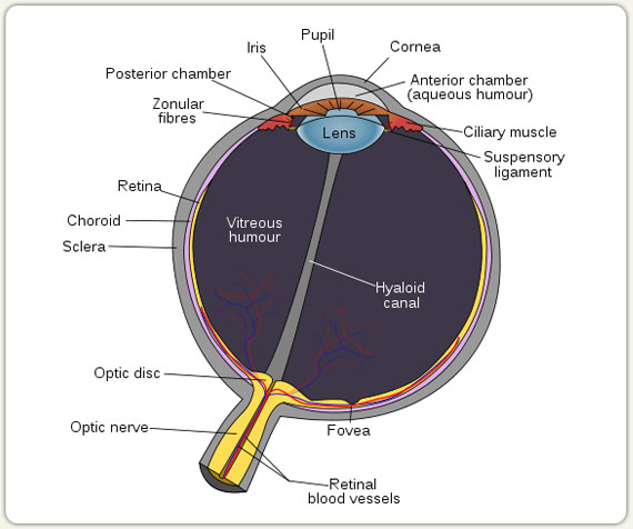
2. LENS?
Your lens is a transparent structure the size and shape of an M & M candy. It is suspended by thin strands, called ZONULES, from a structure called the CILIARY MUSCLE, which runs around your eye immediately behind your IRIS.
Your lens has an inner, tougher center, called the NUCLEUS; a softer outer layer, called the CORTEX; and a skin, called the CAPSULE. When you want to focus up close, your ciliary muscle contracts, and your lens gets fatter; when this muscle relaxes, your lens gets thinner, and your eye focuses at distance.
Your lens grows all through your life, adding one layer each year.
As your lens gets larger, it gets stiffer, and harder to focus. By the time your are in your forties you will no longer be able to change the focus of your lens, so you will need magnifying glasses to read.
A cloudy lens is called a CATARACT.
3. IRIS?
Your IRIS is the part of your eye that gives it color. It is a muscle that has the shape of a lifesaver. Your iris acts like a window shade, opening and closing automatically to change the size of your PUPIL, the black opening in its center. This regulates the amount of light that is admitted into your eye.
The color of your iris-brown, blue, or green-is determined by the amount and distribution of a pigment, MELANIN. Brown eyes have more melanin than blue eyes, so they are less sensitive to bright light.
4. SCLERA?
Your SCLERA is the tough white outer covering of your eye.
5. CONJUNCTIVA?
Your conjunctiva is a flexible, clear mucous membrane that covers the inside of your lids, and the outside of your sclera. Hair-thin arteries and veins run through it. If these blood vessels become inflamed, you have CONJUNCTIVITIS.
Your conjunctiva keeps your eye moist. It is studded with GOBLET CELLS, which produce mucous, an important part of your tears.
6. TEAR FILM?
Tears protect, clean and lubricate your eye. All normal tears have three layers: an outer layer of OIL, which prevents your tears from evaporating; an inner layer of MUCOUS, which keeps your tears attached to your eye; and a middle watery layer, which is complex and fascinating. This middle tear layer is similar to your blood, but without cells. It contains most of the substances found in blood, including salts, sugar, hormones, and antibodies that protect against invading organisms. It is especially high in Vitamin C.
Most of your middle tear layer is produced by your LACRIMAL GLANDS, bean-sized glands which lie just above each upper eyelid. The oil in your tears is made by little bottle-shaped glands, called MEIBOMIAN GLANDS, which are found along the margins of all four eye lids..
7. AQUEOUS?
AQUEOUS circulates steadily through the ANTERIOR CHAMBER of your eye, night and day, bringing oxygen and sugar to your cornea and lens, and washing away wastes. This clear fluid is produced by your CILIARY BODY, which runs around the eye just behind the iris, near the ciliary muscle.
Aqueous drains out of your eye through the TRABECULAR MESHWORK, into SCHLEMM’S CANAL and then into the veins around your eye.
Normally, inflow equals outflow, so the pressure inside your eye is maintained between 8 and 22 millimeters of mercury. However, if your eye has a drain that is not working properly, the pressure inside your eye will rise. This high pressure will eventually damage your OPTIC NERVE, and you will start to go blind. This is “GLAUCOMA”.
8. VITREOUS?
The VITREOUS is the transparent jelly that fills the POSTERIOR CHAMBER of your eye. The vitreous is firm and clear when you are young. As you get older, it gets less firm, and more watery. It eventually collapses on itself, like a tent folding, or a balloon that has lost its air. Because this process detaches the vitreous from the RETINA, it is called a VITREOUS DETACHMENT. After your vitreous detaches you will notice strands called FLOATERS.
9. RETINA?
Your RETINA is an extension of your brain. It is a light-sensitive nerve tissue that lines the inside of your eye, like the film inside a camera. It has 12 layers.
When light is focused by the cornea and lens onto your retina, millions of tiny PHOTORECEPTORS give off electrical signals. These signals feed into 1.2 million OPTIC NERVE fibers. The optic nerve acts as a cable, carrying this information up to the VISUAL CORTEX, which is in the OCCIPITAL LOBE, in the back of your brain.
Most human blindness is caused by diseases of the retina: in infants, amblyopia, or lazy eye; in adults, diabetic retinopathy; in the elderly, macular degeneration.
10. RODS AND CONES?
Photoreceptors come in two different types, 120 million RODS, and 6 ½ million CONES.
Your rods, mainly located in the periphery of your retina, are super sensitive. They operate only when the light is dim. They can only detect large forms and shapes, in black, white, and shades of gray.
Cones, located mainly in the center of the retina, operate in bright light. The ones in your MACULA enable your eye to see details. There are three different types of cones: red, green, and blue. This allows you to have color vision.
11. RETINAL PIGMENT EPITHELIUM?
The retina sits on top of a layer of darkly pigmented cells called the RETINAL PIGMENT EPITHELIUM, or RPE. The RPE is packed with melanin, which absorbs scattered and reflected light. The RPE acts as a nurse for the retina, trimming off the aging tops of the photoreceptors every day, and bringing the retina the chemicals it needs for vision.
12. MACULA?
The MACULA is a tiny area right in the center of your retina. Although it is only the size of the head of a pin, it is responsible for all of your detailed sight. 90% of the signals that go to your occipital lobe come from your macula.
If your macula is damaged, you will no longer be able to read, or drive, or recognize faces. You will, in fact, be legally blind, although you will still retain your peripheral vision.
The little indentation in the very center of your macula is called your FOVEA. The fovea provides your most detailed sight. There are no blood vessels, and no rods, in the fovea.
13. THE BLOOD SUPPLY OF YOUR RETINA?
The inner third of your retina is nourished by your CENTRAL RETINAL ARTERY (CRA) and CENTRAL RETINAL VEIN (CRV). These enter and leave your eye at the OPTIC DISK, the place where the optic nerve originates. They fork, like the branches of a tree, as they run out to the periphery of the retina.
The outer two thirds of your retina is nourished by your CHOROID, a thick layer of blood vessels sandwiched between your RPE and your sclera.
The retina and the conjunctiva are the only parts of your body where your doctor can see your blood vessels. When a doctor looks at these blood vessels, he gets a lot of information about the health of your body.
14. THE EYE MUSCLE SYSTEM?
There are six tiny muscles connected to each of your eyes. They are the size of thick rubber bands. They pull the eye up and down, left and right, and clockwise and counterclockwise.
These muscles are commanded and coordinated by three nerves, located in the brain stem.

